Dicom Viewer Mac Free Download
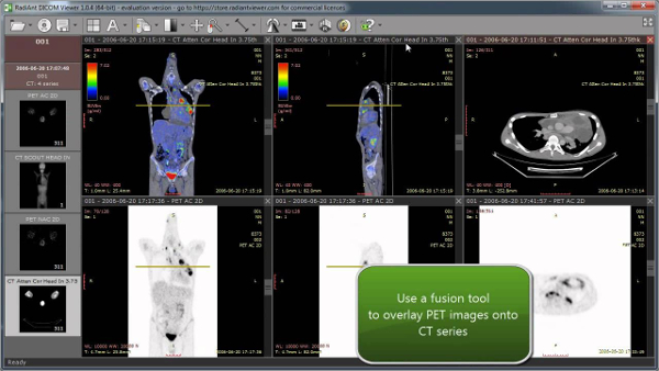
- RadiAnt DICOM Viewer 2020.2 has been available for download for some time now. This versions brings DSA (Digital Subtraction Angiography) to the table. There is an option to set the precise WL/WW values (or SUVbw for PET studies) and to easily create own windowing presets (in 2D, 3D MPR and 3D VR viewers).
- Download dicom viewer for free. Design & Photo downloads - 3DimViewer by 3Dim Laboratory and many more programs are available for instant and free download.
- Syngo fastView 1 is a standalone viewer for DICOM 2 images provided on DICOM exchange media. It can be used on any Windows PC - not allowed on medical workstations. Its operating concept is based on the easy-to-use syngo philosophy; Learn one - know all.
- Microsoft Dicom Viewer Free Download
- Centricity Dicom Viewer Mac Free Download
- Pc Autoplay Dicom Viewer Free Download Mac
- Dicom Image Viewer Free Download
Dicom viewer free download - DICOM Viewer, Free DICOM Viewer, MILLENSYS DICOM Mini Viewer, and many more programs. Enter to Search. My Profile Logout.

syngo fastView Patient Browser:
- Display of patient list from DICOMDIR
- Load via drag&drop or double-click
- Display images from different studies or examinations
syngo fastView Viewer:
- Accurate display of images with syngo graphic objects
- Layouts: 1:1, 4:1, 16:1, 1:2 vertical, 9:1 stripe and stack modes
- Special ultrasound movie mode for cardiac stress echo examinations
- Special 3 D MPR mode for displaying volume datasets in 3 orthogonal views
- Special Fusion mode for displaying resampled fusion datasets side by side
- Simultaneous display of multiple datasets as interactive or automatic movie
- Realtime display in movie mode for multiframe datasets and dynamic MR series
- Implicit load of XA scenes via navigating in movie dialog
- Display of XA scenes with corresponding ECG waveforms
- Display of Digital Subtracted Angiographie images with OR without anatomical background
- Zoom/Pan, Magnify, Minimize, Magnifying Glass, Magic Glass (Capture Area)
- Rotate, Flip, Mirror, Home View
- Windowing; Standard Window, Window 1 and 2
- Modality-specific image text (configurable)
- Save images in JPEG, BMP or AVI formats
- Direct e-mail access with image converted in JPEG format
- Print out images on all installed printers
- Multi-Language support (English, German, French, Spanish, Chinese, Japanese)
- Easy access to standard functions via TaskCard icons
- Dialog for displaying DICOM attributes of selected image
View Your Own Medical Scans in 3D on Mac
Load your DICOM files from CD’s, USB’s and local directories and interact with your own anatomy in fully-immersive 3D.
MacOS Certified Application for easy installation & viewing.
Free DICOM Viewer For Mac With Full 3D Visualisation
We’ve optimised our proprietary Volume Rendering Platform to work on your MacOS device and convert (render) your private medical files locally on your computer to ensure that your confidential data stays with you. 3Dicom For Mac works on MacOS however we’re working on an iOS version for iPad & iPhone, launching soon!
Mac Multi-Touch
Our MacOS version includes support for Apple Multi-Touch, allowing for simple, intuitive rotation, panning and zooming using multiple fingers on your TouchPad.
View In Both 2D & 3D
Scroll through your individual orthogonal planes in the 2D views to identify particular structures and then view their location in 3D space with the interactive 3D view.
Compatible with CT/MRI/PET
Load in almost any ‘slice’ based DICOM image such as Computed Tomography (CT), Magnetic Resonance Imaging (MRI) or Positron Emitting Tomography (PET).
Capture Screenshots In 3D
Easily capture screenshots in the full 3D view with the QuickCapture tool. Common JPG file format for easy post-processing & sharing.
Loads In 90s or less
Typical DICOM scans load within 90 seconds or less using just your computer’s GPU, no internet connection required at all.
Optimised Graphics
The MacOS version has been optimised to provide the best possible trade off between processing/navigation speed and resolution.
Common Use Cases for 3Dicom Viewer for MacOS
In line with our vision of developing better health literacy, we’re always pleased to hear from users and how they’ve used 3Dicom Viewer and the Volumetric Rendering Platform developed by Singular Health Group for improved health outcomes and to empower more informed discussions with their healthcare professionals.
Improved Spatial Understanding of Anatomical Structures
We’ve taken the complex task of finding the right Hounsfield Units to show your required tissue density and simplified it into an intuitive slider. With our Windowing Values sliders, you can very quickly “strip” away different densities to expose particular areas of interest.
As seen in the image, increasing the lower threshold of the Hounsfield Units progressively removes the softer tissue, allowing you to view the internal structures such as cartilage, muscle, organs and ultimately the high-density skeletal system.
Adjusting your windowing should be the first step once your scan has loaded prior to editing the Display Settings or using the 3D slicer tool.
Microsoft Dicom Viewer Free Download
Viewing Pathology in 3D
Our lightweight 3D viewer isn’t intended as a full clinical DICOM viewer, rather as a fast and easy way to review your 2D analysis in the 3D space. Your time is precious and despite using your own computer’s processing power to render the dicom (.dcm) or NiFti (.nii) files into a 3D volumetric render, we’ve optimise our conversion and rendering process to take less than 60 seconds.
In fact, most scans load in less than 30 seconds, giving you a quick-fire tool to create, review and capture (via screenshot or screen recording) a better understanding of complex cases.
Screenshots of 3D Rendered Anatomy for Presentations & Education
Utilising the Display setting sliders in the control panel, you can manipulate the Brightness, Contrast and Opacity values to see more in your 3D render. Simulate an ‘X-Ray’ view by reducing the opacity value and get a wire-frame type view that really highlights particular areas (as seen with the vascular system in the image).
Due to the way in which Hounsfield Units (tissue density/windowing) uses grey-level mapping which impacts upon brightness and contrast, ensure that you try editing these two values after finding your desired Windowing range. This will help accentuate your scan without changing the accuracy of the medical scan data.
Immersive Exploration of Patient Anatomy
Get up close and personal with your scans. We’ve built in a Zoom feature that is easily controlled by your Up & Down arrow keys to zoom in and out and get the best position for capturing images.
Soon, you’ll be able to voyage inside the 3D model using our Immersive Zoom tool, positioning the 3D rendered model and navigating into internal spaces such as the trachea, lungs and more.
How To Optimise Your Scan in 3Dicom Viewer for Mac OS
Follow the 3 easy steps below to load and enhance your medical scans.
Load and Convert Your Dicom Files
We’ve taken the complex task of finding the right Hounsfield Units to show your required tissue density and simplified it into an intuitive slider. With our Windowing Values sliders, you can very quickly “strip” away different densities to expose particular areas of interest.
As seen in the image, increasing the lower threshold of the Hounsfield Units progressively removes the softer tissue, allowing you to view the internal structures such as cartilage, muscle, organs and ultimately the high-density skeletal system.
Adjusting your windowing should be the first step once your scan has loaded prior to editing the Display Settings or using the 3D slicer tool.
Enhance the converted 3D model
Get up close and personal with your scans. We’ve built in a Zoom feature that is easily controlled by your Up & Down arrow keys to zoom in and out and get the best position for capturing images.
Soon, you’ll be able to voyage inside the 3D model using our Immersive Zoom tool, positioning the 3D rendered model and navigating into internal spaces such as the trachea, lungs and more.
Explore & Analyse The 3D Anatomy
As seen in the video, now that you’ve focused in on the most relevant anatomy and enhanced the image with improved brightness and contrast, you can view it from all angles.
Using the Shift Key + your mouse pad, you can pan (move side to side) the view point.
To rotate the 3D volumetric model, simply click and drag.
Finally, using a combination of panning, rotating and then moving/zooming through the model, you can actually position yourself inside your body and explore it from the inside out!
Introducing 3Dicom Pro for MacOS
Impressed by the 3Dicom Viewer but looking to get even more information from your DICOM medical images? 3Dicom Pro is the culmination of thousands of hours of work developing many industry-standard tools optimised for speed and intuitive use within our cross-platform software.
For just US$5/month you can access many additional features going beyond visualisation & allowing for in-depth analysis of radiological scans.
View Dicom Meta Data
3Dicom Pro has a number of different processes by which to read and interpret the various DICOM standards so you’re able to read and filter all DICOM meta-tags.
Additional Presets
From bony and soft-tissue windowing through to auto-contrast and brightness settings, presets accelerate the process of viewing regions of most interest.
Centricity Dicom Viewer Mac Free Download
Hounsfield Histogram
Pc Autoplay Dicom Viewer Free Download Mac
Extracting the precise Hounsfield (density) values from the DICOM meta-data, we enable you to better analyse and threshold your scans and manually set upper & lower values.
Save Measurements
Save your measurements and annotations and attach them to a new DCM file for sending to medical practitioners.
Convert DICOM to PNG
Dicom Image Viewer Free Download
Save each DICOM slices as PNGs along with screen captures and videos of the 3D volume rendered output. PNG’s capture markups too!
2D & 3D Measurements*
Add length and angle measurements to 2D slices and watch as the resultant 3D vector (length) is generated in the 3D model.
And much more….
